


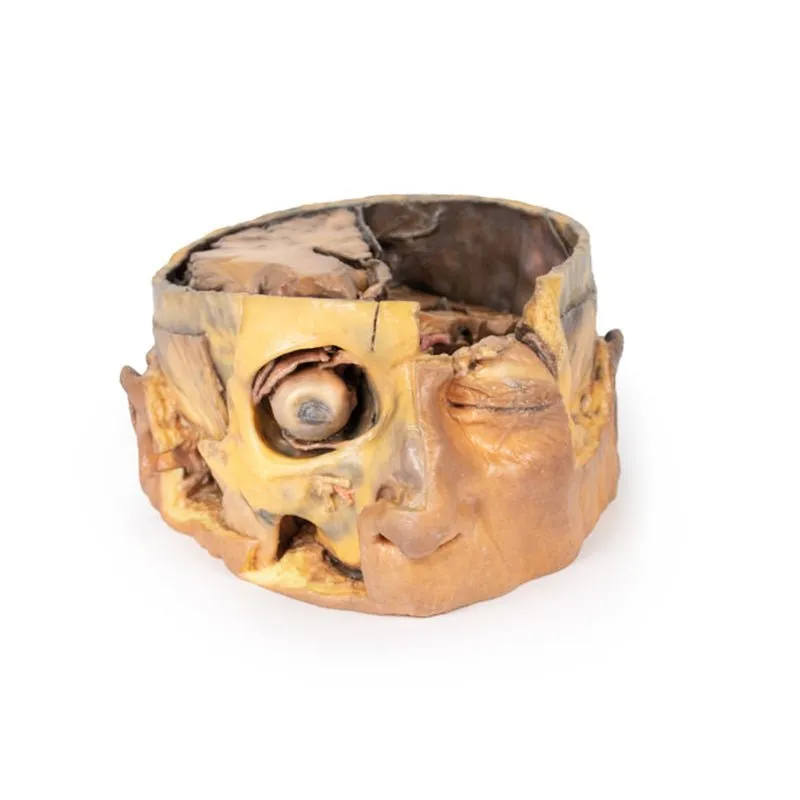
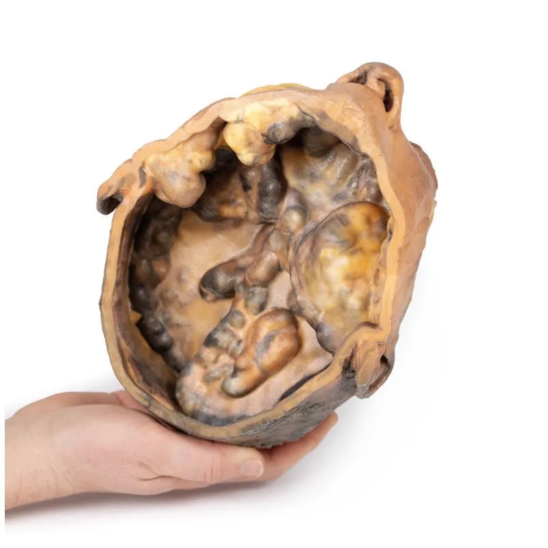
3D Printed Transverse Section of the Head
This 3D model preserves a transverse section through the cranial cavity with partial dissection of the brain and
exposure of the left orbital roof, alongside a deep dissection of the face and temporomandibular joint
region.
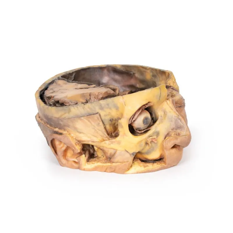
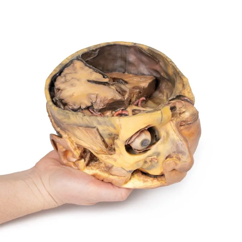
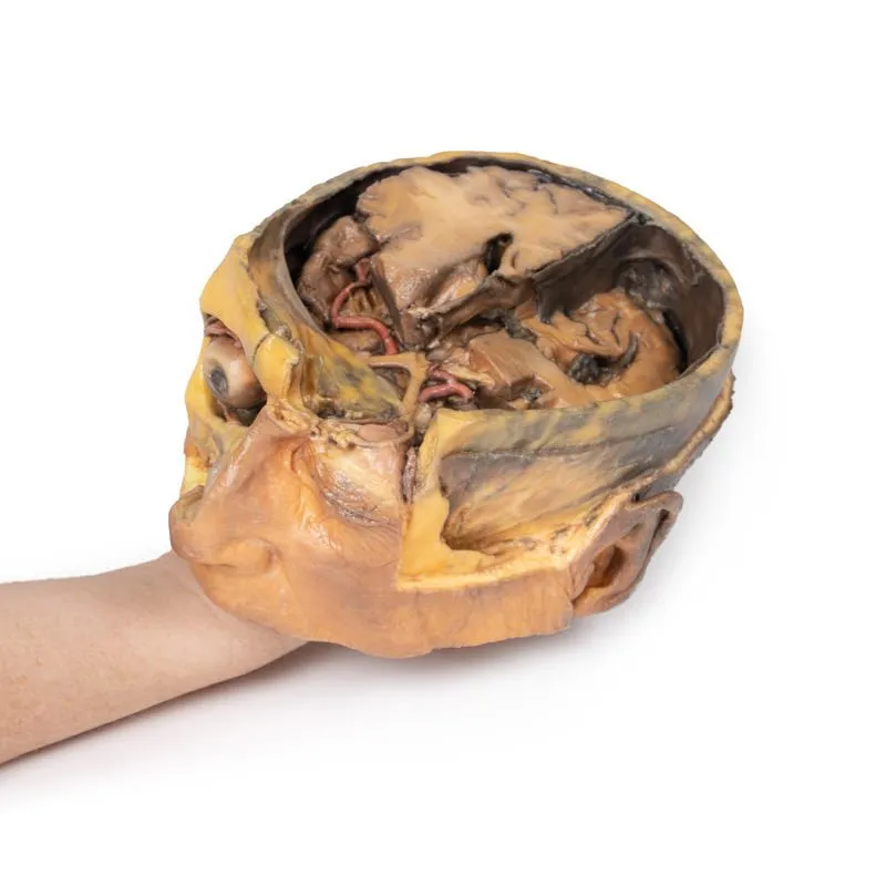
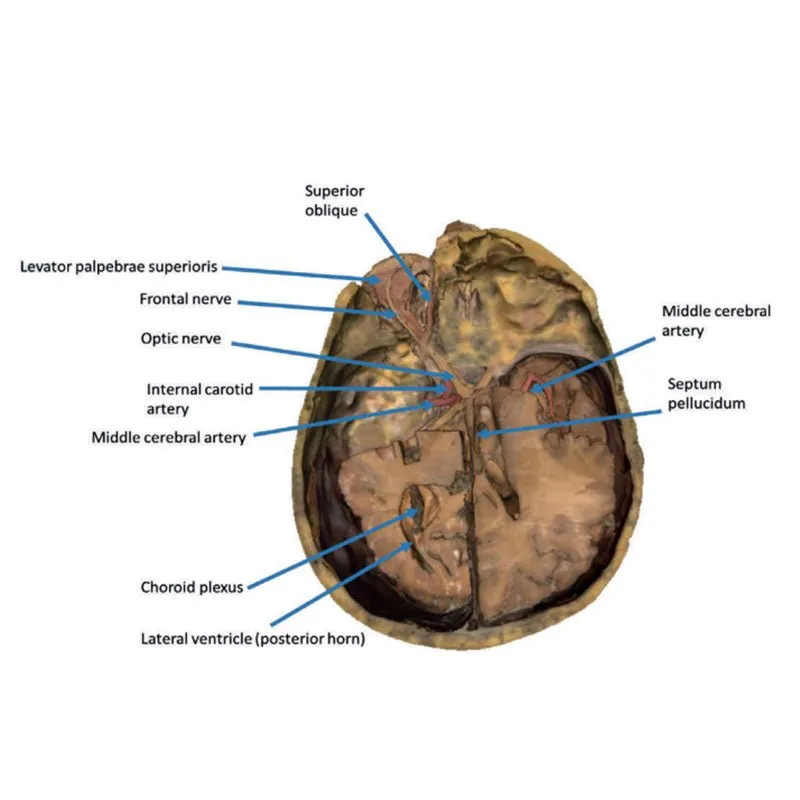
Within the cranial cavity, the dura mater has been largely removed from the anterior cranial fossa, with
retention of the layer in part across the middle and posterior cranial fossae. On the right side, the cerebrum has
been dissected to expose the lateral ventricle and to open the lateral fissure to demonstrate the course of the
middle cerebral artery between the frontal, parietal and temporal lobes. A more significant dissection of the brain
on the left side allows for an appreciation of the midline third ventricle and retained septum pellucidum on the
right side, the falx cerebri (with the superior sagittal sinus visible in cross-section), and parts of the anterior
and posterior horns of the lateral ventricle with choroid plexus. This differential dissection of the brain also
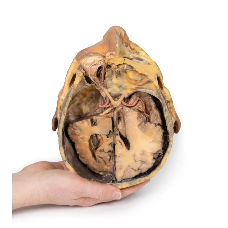
provides an excellent view of the optic nerves, chiasm and tracts, and the relation of these nervous structures to
the left internal carotid artery, and bases of the anterior and middle cerebral arteries.
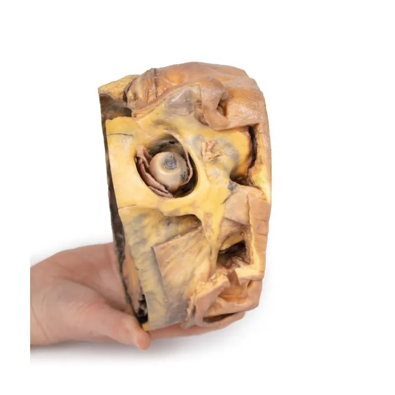
Anteriorly in the
cranial cavity, the left optic nerve can be followed into the left orbit, which has been opened to expose several
key orbital structures. Centrally, the frontal nerve is well-preserved on the levator palpebrae superioris muscle.
Laterally, the lacrimal gland rests in the superior quadrant, while medially the partial dissection into the frontal
bone and sinuses affords a clear view of the superior oblique muscle passing through the trochlea. Deep to these
superficial structures extraocular fat has been removed to show the medial rectus muscle laterally, and the
nasocilliary nerve and medial rectus muscles medially.
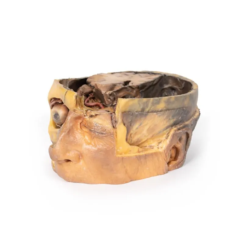
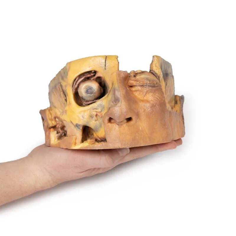
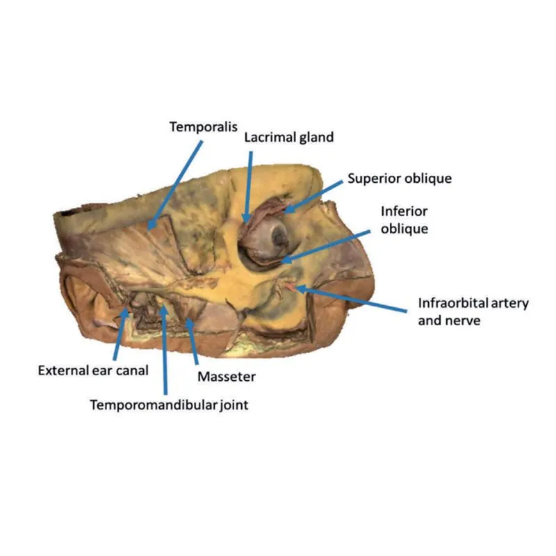
In the face, the skin, superficial tissue, orbicularis
oculi and extraocular fat have been removed from the right orbit to expose the extraocular muscles and lacrimal
gland. The levator palpebrae superioris is well-defined despite being detached from the superior tarsal plate. The
reflection of the superior oblique muscle from the trochlea onto the eye can be seen, as well as the insertions of
the medial and lateral rectus muscle, and the full course of the inferior oblique.
Across the rest of the right
side of the face and temporal region a deep dissection has exposed a number of structures. Inferior to the orbital
margin, the infraorbital artery and nerve have been exposed exiting via the infraorbital foramen. The superficial
and deep heads of the masseter are well defined, and the partial dissection of the temporalis muscle provides a
perspective on its broad origin and depth of fibres near pterion (and in contrast to the exposed and undissected
right side). Inferior to the zygomatic, the parotid gland has been dissected to expose the mandibular condyle
resting in the glenoid fossa and to demonstrate the relationship of the external ear relative to the external
auditory meatus.
GTSimulators by Global Technologies
Erler Zimmer Authorized Dealer


